
Neural circuits – Understanding the networks of the brain and nervous system
Welcome to Neural Circuits, a site dedicated to the study of neural circuits: anatomy, function, role in behaviors, research techniques, and applications in neuroscience.

Neuronal Physiology
Nervous tissue is primarily composed of two types of cells: neurons and glial cells.
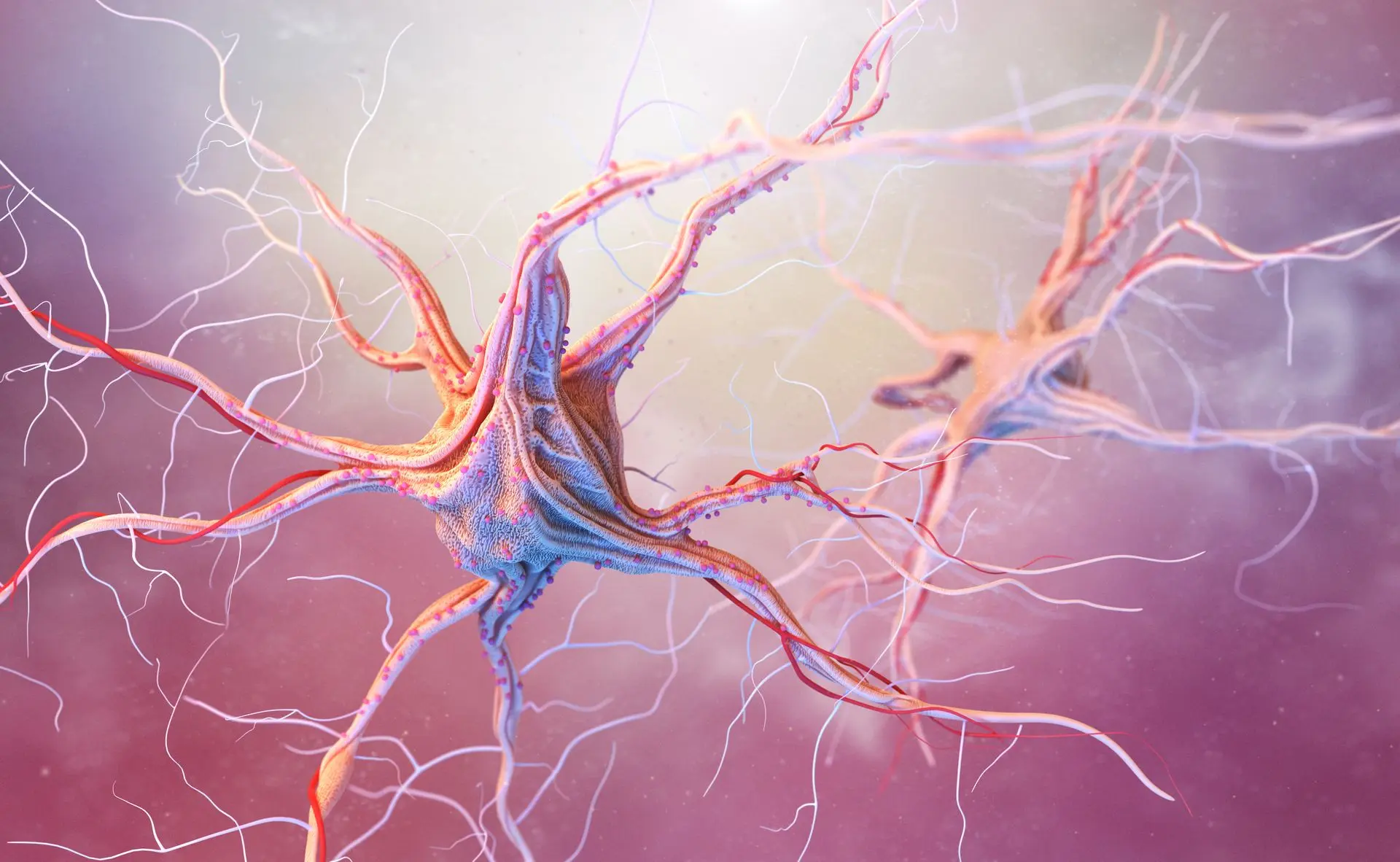
The neuron is the fundamental structural and functional unit of the nervous system. It is a highly specialized and differentiated cell responsible for conducting nerve impulses. The neuron consists of a cell body from which two types of processes extend: dendrites and an axon. These processes form the nerve fibers, which may be surrounded by a protective covering called the myelin sheath a fatty, white substance that insulates and enhances signal transmission.
Types of Neurons
Structural Classification:
| Functional Classification of Neurons :
|

Glial Cells
Glial cells form a tissue closely associated with neurons. They provide structural support and insulation within the central nervous system (CNS), fulfilling functions similar to connective tissue including support, exchange, and nutrition.
In the CNS, there are four main types of glial cells:
- Astrocytes: These cells play a key role in nourishing neurons. They have numerous branching processes that extend in all directions, sending projections toward blood vessels to facilitate exchange between blood and nervous tissue.
- Oligodendrocytes: Found in the CNS, these cells are responsible for producing myelin and exhibit rhythmic movement.
- Microglial cells: These are the macrophages of nervous tissue; they remove waste and debris. They can migrate as needed to sites where cellular debris must be cleared.
- Ependymal cells: These are epithelial-like cells that form the lining of the brain’s ventricles and the central canal of the spinal cord.
In the Peripheral Nervous System (PNS), two types of glial cells are found:
- Schwann cells (neurolemmocytes): involved in the production of myelin in the PNS.
- Satellite cells: provide support and regulate the environment around neuron cell bodies in ganglia.

Neural Impulse Transmission
Just like all cells in the body, the neuron's membrane is polarized: the exterior is positive, and the interior is negative. This polarity is due to a concentration gradient of sodium (Na⁺) and potassium (K⁺) ions on either side of the plasma membrane. At rest, this gradient is maintained by the Na⁺/K⁺ ATPase pump, resulting in a resting potential of about -70 mV.
When the membrane's permeability to certain ions changes momentarily, ion exchanges occur across the membrane, leading to depolarization. If the depolarization reaches a sufficient level, it triggers an action potential a wave of depolarization that travels along the axon. This traveling depolarization constitutes the nerve impulse.
Action Potential
An action potential is a characteristic of excitable cells and involves a rapid depolarization followed by repolarization of the membrane. It occurs in four key phases:
1️⃣ Depolarization (rising phase)
After stimulation, voltage-gated sodium channels open rapidly, allowing sodium ions to rush into the cell. The membrane potential moves towards sodium’s equilibrium potential (+65 mV), though it doesn’t fully reach it.
2️⃣ Repolarization
Sodium channels close, and voltage-gated potassium channels open. Potassium exits the cell, restoring the membrane potential towards its resting state.
3️⃣ Hyperpolarization
Potassium conductance remains elevated for a short time, causing the membrane potential to dip below its normal resting level (around -75 mV).
4️⃣ Return to Resting Potential
The Na⁺/K⁺ ATPase pump restores the original ion distribution, pushing Na⁺ out and bringing K⁺ back in. This enables the action potential to propagate along the neuron, forming the physical basis of the nerve impulse.
Action Potential Properties
🔹 Threshold: The minimum stimulus strength required to trigger an action potential.
🔹 All-or-nothing law: Once threshold is reached, the action potential occurs fully, regardless of stimulus strength.
🔹 Summation:
- Temporal summation: Two sub-threshold stimuli close in time can combine to trigger an action potential.
- Spatial summation: Two sub-threshold stimuli from nearby locations can combine to trigger a response.
Refractory Periods
🔸 Absolute refractory period: No action potential can be triggered, no matter the stimulus strength (Na⁺ channels are inactivated).
🔸 Relative refractory period: A stronger-than-normal stimulus can trigger an action potential as the membrane returns to rest.
➡ These periods ensure the impulse only travels in one direction along the neuron.
Nerve Impulse Conduction
🔹 Continuous conduction
In unmyelinated fibers, depolarization occurs progressively along the axon. Each sodium channel opens in sequence, spreading the impulse.
🔹 Saltatory conduction
In myelinated fibers, the impulse jumps from one node of Ranvier to the next, greatly increasing the speed of conduction.
The nerve impulse is transmitted to another neuron or an effector cell (e.g., muscle, gland) at a specialized junction called a synapse.
👉 Types of synapses
- Electrical synapse: Direct transmission of impulses through gap junctions (rare in mammals).
- Chemical synapse: Involves neurotransmitters crossing a synaptic cleft between the presynaptic and postsynaptic membranes.
👉 Components of chemical synapse
- Presynaptic membrane: Contains neurotransmitter-filled vesicles.
- Synaptic cleft: Gap between the two cells.
- Postsynaptic membrane: Contains receptors for the neurotransmitter.

How a Chemical Synapse Works
1️⃣ The action potential reaches the axon terminal, opening voltage-gated calcium channels.
2️⃣ Calcium enters the presynaptic neuron, triggering exocytosis of neurotransmitters into the synaptic cleft.
3️⃣ The neurotransmitter binds to receptors on the postsynaptic membrane, altering ion permeability and membrane potential.
➡ Inhibitory neurotransmitters (e.g., GABA) open chloride channels, hyperpolarizing the postsynaptic membrane.
➡ Excitatory neurotransmitters (e.g., glutamate, acetylcholine) open sodium channels, depolarizing the membrane and possibly triggering an action potential.
Cell Culture and Synapse Formation In Vitro
What is synapse culture?
Cell culture of synapses refers to laboratory techniques that allow neurons to grow and form functional synaptic connections outside of the organism, in a controlled artificial environment (in vitro). These models are fundamental for studying the molecular, electrical, and biochemical mechanisms of synapse formation, function, and plasticity.
🔬 Why culture synapses?
Synaptic cell culture provides:
- A simplified and controlled environment to study neuronal development and synaptic connectivity
- The ability to observe synapse formation, maturation, and elimination
- A platform for pharmacological testing (e.g., drugs targeting synaptic transmission)
- Insight into synaptic defects in neurological diseases (e.g., autism, epilepsy, neurodegeneration)
🧠 Common sources of neurons for synapse culture
- Primary neurons: harvested from embryonic or neonatal rodent brains (e.g., hippocampus, cortex, spinal cord)
- Human iPSC-derived neurons: induced pluripotent stem cells reprogrammed into neurons, allowing study of human synapses
- Neuroblastoma or hybridoma lines: used in simplified or large-scale assays
🧪 Techniques in synaptic cell culture
1️⃣ Substrate preparation
- Neurons are cultured on coated coverslips or culture dishes.
- Common coatings: poly-D-lysine, laminin, collagen → promote adhesion and neurite outgrowth.
2️⃣ Growth media
-
Specialized neurobasal or serum-free media supplemented with:
- B27 or N2 supplements
- Glutamine
- Antibiotics
- Growth factors (e.g., BDNF, NGF) in some protocols
3️⃣ Synapse formation
- After a few days in culture, neurons extend axons and dendrites.
- Synapses begin forming at contact points where axons meet dendrites (visualized with fluorescent markers like synapsin, PSD-95).
-
Functional synapses are confirmed by:
- Calcium imaging
- Patch-clamp electrophysiology
- FM dye uptake/release assays
4️⃣ Co-culture systems
- Neuron-glia co-cultures: Astrocytes or oligodendrocytes support synapse maturation and maintenance.
- Neuromuscular co-cultures: Neurons form synapses on muscle fibers → used for studying neuromuscular junctions.
⚡ Applications of synaptic cell culture
- Study of synaptic plasticity (e.g., long-term potentiation or depression)
- Modeling neurological diseases (e.g., Alzheimer’s, schizophrenia)
- High-throughput screening of synaptotoxic or neuroprotective compounds
- Investigation of synaptic development and pruning
- Analysis of gene editing effects (e.g., CRISPR-modified neurons)
🧫 Example: Synapse imaging in culture
-
Immunostaining for:
- Presynaptic markers: Synapsin, synaptophysin
- Postsynaptic markers: PSD-95, Gephyrin
- Confocal microscopy or super-resolution imaging helps visualize and quantify synaptic contacts.
🌟 Advantages of synapse culture
✅ Direct visualization of synaptic structures
✅ Fine control over experimental variables
✅ Accessibility for genetic manipulation (viral vectors, transfection)
✅ Ethical alternative to animal models for certain studies
Access the full scientific article in PDF by clicking here.